
Executive Key Takeaways
Executive Summary: Shockwave therapy (ESWT) is a structured, non-surgical option for chronic tendon and fascia pain in San Diego that works by stimulating tissue repair and improving load tolerance rather than providing immediate “numbing” relief. The best outcomes occur when the correct pain generator is confirmed on exam and ESWT is paired with progressive, condition-specific strengthening between sessions.
- Diagnosis-First Candidate Selection: High-quality clinics confirm a clearly reproducible tendon/fascia pain source through orthopedic testing (palpation, ROM/strength measures, functional provocation) and use imaging only when it changes management.
- Protocol-Driven Treatment and Rehab Pairing: Most plans use 3–6 sessions spaced about 5–10 days apart, with dosing adjusted to tissue depth and irritability while a graded loading program (eccentric or heavy-slow resistance) drives long-term capacity gains.
- Expected Timeline and Safe Activity Rules: Improvement typically accumulates over weeks as remodeling occurs, with common 24–72 hour post-treatment soreness requiring temporary impact reduction while continuing guided mobility and strengthening.
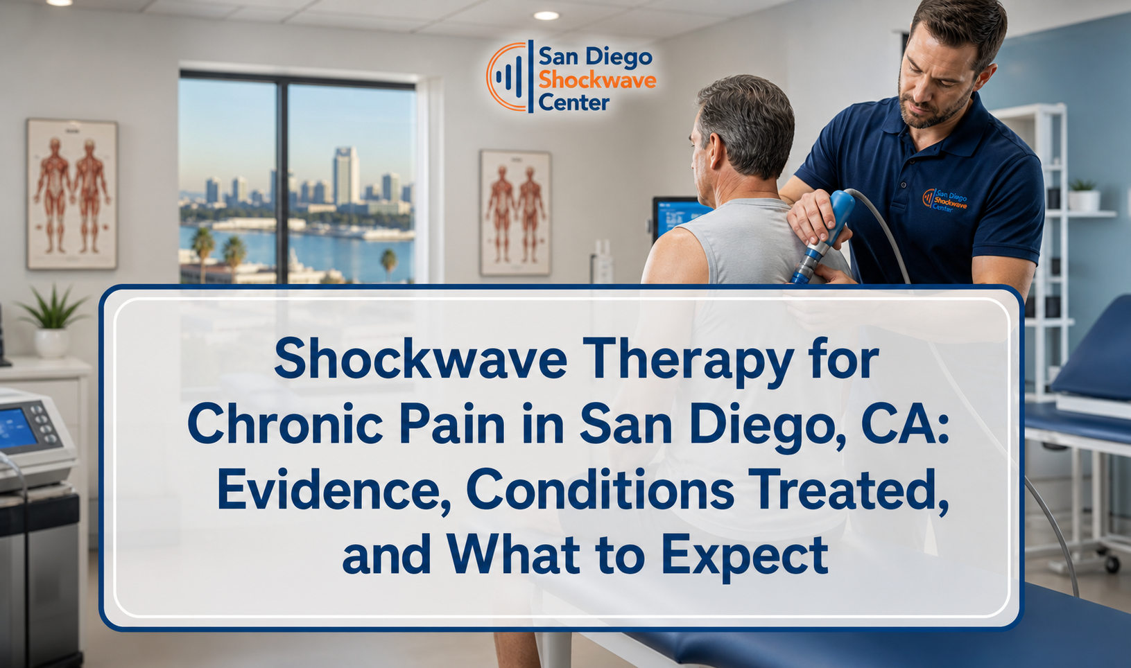
Shockwave therapy is a non-surgical treatment that uses focused acoustic pressure waves to stimulate tissue repair and reduce chronic musculoskeletal pain. Shockwave Therapy for Chronic Pain San Diego commonly targets stubborn conditions like plantar fasciitis, Achilles tendinopathy, patellar tendinopathy, tennis elbow, shoulder calcific tendinopathy, and greater trochanteric pain syndrome. Clinicians in San Diego typically confirm candidacy with a detailed orthopedic exam, palpation of the pain generator, range-of-motion and strength testing, and functional checks like heel-rise tolerance, single-leg squat control, or grip pain provocation. Imaging review may be used when needed, including ultrasound for tendon thickening or calcific deposits, and X-ray for calcific shoulder findings. A typical session lasts about 10–20 minutes and applies thousands of pulses to the affected tendon or fascia, with intensity adjusted to tolerance and tissue depth. Most care plans involve 3–6 sessions spaced about 5–10 days apart, combined with load management and progressive rehab exercises such as eccentric calf lowering for Achilles pain or progressive plantar fascia loading for heel pain. Expect localized soreness, redness, or swelling for 24–72 hours, and avoid heavy impact activity during that window while continuing guided mobility and strengthening. Results often build over weeks as collagen remodeling and local circulation improve, rather than delivering instant numbness like an injection.
How shockwave therapy works for chronic tendon and fascia pain
Shockwave therapy (extracorporeal shockwave therapy, ESWT) uses controlled acoustic pulses to create a biological response in chronically overloaded tissue rather than masking symptoms. The objective is to restart a stalled healing cycle by improving local circulation, modulating pain signaling, and supporting collagen remodeling.
In chronic tendinopathy and fasciopathy, tissue often shows impaired load tolerance, disorganized collagen, and pain with specific functional tasks (jumping, gripping, stairs, first-step pain). Focused or radial shockwave is applied over the most symptomatic tissue and nearby attachment zones to stimulate a repair response. Clinically, this is treated as a load-management and tissue-capacity problem—not an “inflammation-only” issue—so a progressive strengthening plan remains essential.
- Mechanotransduction: pressure waves create mechanical signaling that influences cellular activity (including fibroblast behavior and collagen organization).
- Neuro-modulation: therapy can reduce pain sensitivity in the treated region, which can improve tolerance to rehab loading.
- Local perfusion changes: improved microcirculation is a common treatment goal in chronically painful tendon and fascia tissue.
For a plain-language definition of extracorporeal shockwave therapy and its general medical use, see extracorporeal shockwave therapy.
Who is a good candidate in San Diego (and who is not)
Appropriate candidates have a clearly reproducible musculoskeletal pain generator (tendon, fascia, or enthesis) that has not improved with well-executed conservative care. Contraindications and precautions are screened using standard outpatient orthopedic intake and risk checks.
Clinicians typically look for a consistent pattern: localized tenderness on palpation, pain provocation with resisted testing (e.g., wrist extension for lateral epicondylalgia), and functional limitation (stairs, running, lifting). The most predictable outcomes occur when treatment selection matches tissue type and the patient can follow a graded loading plan between sessions.
Common “best-fit” profiles
These cases usually match the clinical indications most often treated with ESWT. They have persistent symptoms, clear mechanical triggers, and no urgent red flags.
- Plantar heel pain consistent with plantar fasciopathy (often “first-step” pain)
- Achilles tendinopathy (mid-portion or insertional) with pain during running/jumping
- Tennis elbow (lateral epicondylalgia) provoked by gripping and wrist extension
- Patellar tendinopathy or chronic anterior knee pain patterns with tendon load sensitivity
- Calcific shoulder tendinopathy where calcific deposits are confirmed on imaging
- Greater trochanteric pain syndrome with reproducible lateral hip tenderness and load pain
When shockwave therapy may be inappropriate
Shockwave should not be used when medical risk is unacceptable or when the pain source is not a treatable tendon/fascia target. Screening is not optional—particularly for bleeding risk, pregnancy considerations, and suspected serious pathology.
- Suspected fracture, infection, tumor, or systemic red flags (requires medical evaluation first)
- Open wounds or uncontrolled skin infection at the treatment site
- Pregnancy (avoid treating near the trunk/pelvis; providers commonly defer ESWT)
- Bleeding disorders or anticoagulant therapy (requires individualized medical clearance due to bruising/bleeding risk)
- Unclear diagnosis (diffuse pain without a clear pain generator is a poor match)
What an evidence-aligned visit looks like (exam, imaging, and treatment planning)
A quality shockwave plan is built on diagnosis confirmation and measurable baselines, not “pain-only” reporting. Most San Diego clinics follow a predictable outpatient flow: focused exam, functional testing, and selective imaging review when it changes management.
The provider typically establishes (1) the anatomical pain generator, (2) the movement or load that reproduces symptoms, and (3) a progression strategy that reduces flare-ups while increasing capacity. This is also where expectations are set: the response is commonly gradual over several weeks.
Core orthopedic exam components
These steps identify the pain generator and rule out competing diagnoses. The goal is to make the condition reproducible and measurable before any pulses are applied.
- History: onset, training/workload changes, morning stiffness pattern, prior treatments, medication review.
- Palpation mapping: pinpoint tenderness at tendon insertions (enthesis) or mid-substance, fascia origin, or calcific hotspot.
- Range of motion: joint mobility restrictions that increase tendon load (ankle dorsiflexion limits, hip control deficits, etc.).
- Strength testing: isometric and isotonic testing to identify pain-limited weakness.
- Functional tests: heel-rise tolerance, hop tests (when appropriate), single-leg squat control, grip dynamometry or provocative grip tasks.
Imaging: when it is used (and when it is not)
Imaging is most useful when it changes the plan—such as confirming calcific tendinopathy or excluding other pathology. It is not mandatory for every patient with a classic presentation.
- Ultrasound: may be used to evaluate tendon thickening, neovascularity patterns, partial tears, or calcific deposits.
- X-ray: commonly used when calcific shoulder tendinopathy is suspected, or to assess bony contributors (e.g., heel spurs as contextual findings).
- MRI: reserved for complex presentations, suspected major tears, or when symptoms fail to correlate with exam findings.
Session structure, dosing logic, and what you feel during treatment
A typical session is short (often 10–20 minutes) and delivers thousands of pulses to a targeted area. Intensity is adjusted to tissue depth, irritability, and patient tolerance while maintaining a therapeutically meaningful dose.
Most protocols in outpatient practice use a multi-session plan spaced roughly weekly, because tissue adaptation and collagen remodeling are not immediate. The practical goal is to reduce pain enough to restore progressive loading without provoking prolonged flare-ups.
Typical session workflow
Standardized workflow improves consistency across sessions and helps clinicians re-check progress objectively. Most visits follow a repeatable set of steps.
- Re-test baseline: repeat a consistent measure (first-step pain rating, heel raises to pain, grip pain provocation).
- Mark the target: identify the most symptomatic point and surrounding referral zone.
- Apply coupling gel: improves transmission of acoustic waves.
- Deliver pulses: adjust intensity based on depth and tolerance; avoid “white-knuckle pain” dosing.
- Post-care plan: activity modification for 24–72 hours and specific loading exercises.
What sensations are normal vs. not normal
Temporary local soreness is expected, but severe or escalating symptoms are not the goal. Good clinics explicitly differentiate normal post-treatment discomfort from warning signs.
- Common and expected: localized soreness, redness, mild swelling, tenderness for 24–72 hours.
- Less common: bruising, especially in sensitive areas or when patients bruise easily.
- Stop-and-call situations: severe pain spike that does not settle, new numbness/tingling, signs of infection, or inability to bear weight that is worse than pre-treatment.
For a practical, patient-focused outline of timing, soreness windows, and activity rules, review shockwave therapy recovery basics.
Conditions commonly treated in San Diego and how plans differ by body region
Shockwave is not a one-size-fits-all procedure; the tissue target and rehab loading must match the diagnosis. The best results occur when the provider pairs ESWT with a region-specific strengthening progression and clear return-to-activity rules.
Below are common chronic pain targets and how clinicians typically structure the “between-session” work so the tendon or fascia adapts rather than flares.
Plantar fasciitis / plantar fasciopathy
Heel pain plans focus on reducing morning pain and restoring foot-ankle load tolerance. Loading is progressed from controlled isometrics to heavier calf/foot strengthening while avoiding sudden impact spikes.
- Common triggers: first-step pain, prolonged standing, rapid increases in walking/running volume.
- Rehab pairing: plantar fascia-specific loading, calf strengthening, foot intrinsic work, step-down control.
- Activity rule: avoid new plyometrics during the 24–72 hour soreness window after treatment.
Achilles tendinopathy
Achilles plans emphasize graded calf loading and controlled return to running. Dosing decisions often differ for mid-portion vs. insertional pain because compression sensitivity and ankle dorsiflexion tolerance can vary.
- Rehab pairing: heavy slow resistance or eccentric-biased calf work, soleus capacity training, hip control for gait mechanics.
- Monitoring metric: pain during/after calf raises and the next-morning response.
Tennis elbow (lateral epicondylalgia)
Elbow plans target extensor tendon load tolerance and grip capacity for work, lifting, or sport. Shockwave is typically combined with progressive wrist extensor strengthening and graded grip exposure.
- Rehab pairing: isometric then isotonic wrist extension, supination/pronation control, grip progression.
- Behavioral modification: reduce repetitive high-force gripping temporarily; adjust tools/ergonomics.
Chronic knee pain (patellar tendon and related anterior knee patterns)
Knee protocols aim to reduce tendon sensitivity and restore squat, stair, and jump tolerance. Load is progressed carefully to avoid symptom rebound that can occur with aggressive return-to-sport timelines.
- Rehab pairing: quad strengthening (isometrics to heavy slow resistance), hip abductor strength, tendon-friendly squat progressions.
- Functional benchmark: controlled single-leg squat and step-down without sharp tendon pain.
If knee pain is your primary limiter, see Shockwave Therapy for Chronic Knee Pain for a condition-specific overview.
Compulsory reference table: treatment parameters and local care standards
This table summarizes concrete, patient-relevant metrics and the practical “rules” most clinics use to keep treatment consistent and safe. It is designed to help readers compare plans and ask better questions during an intake visit.
| Feature / Metric | Specifications | Local Guidelines |
|---|---|---|
| Typical session length | ~10–20 minutes of on-site treatment time | Allow additional time for re-testing, education, and exercise updates |
| Session count and spacing | Commonly 3–6 sessions, often spaced ~5–10 days apart | Progress is checked with repeatable functional tests before each session |
| Immediate after-effects | Soreness/redness/swelling commonly 24–72 hours | Avoid heavy impact during the soreness window; continue guided mobility/strength work |
| Candidacy confirmation | Orthopedic exam + palpation + ROM/strength testing; imaging as needed | Ultrasound/X-ray review is commonly used for calcific shoulder or uncertain tendon pathology |
| Rehab pairing requirement | Progressive loading plan (e.g., eccentric or heavy slow resistance) | Exercise progression is adjusted based on next-day response, not same-day pain only |
| Outcome timeline | Typically builds over weeks as tissue remodeling occurs | Patients are coached to track function (walking tolerance, stairs, grip) rather than expecting instant numbness |
How to choose a San Diego provider: objective quality checks
Provider selection should be based on diagnostic accuracy, measurable outcomes, and integration with rehab—not marketing claims. You can screen quality quickly by asking about testing, dosing logic, and how return-to-activity decisions are made.
Because ESWT is procedure-based, good clinics document baselines and re-test them. This helps determine whether the plan needs modification (different target zone, different loading plan, or different diagnosis).
- Diagnosis clarity: they can name the structure being treated and reproduce your pain with specific tests.
- Measurable baselines: they use at least one repeatable functional test (heel raises, grip provocation, step-down tolerance).
- Rehab integration: they prescribe a progressive strengthening plan and modify it session-to-session.
- Risk screening: they ask about blood thinners, bleeding disorders, pregnancy, and red flags.
- Activity rules: they give specific post-treatment guidance for the 24–72 hour window.
How to set expectations: realistic outcomes and decision points
ESWT is best viewed as a catalyst for recovery, not a replacement for strengthening or a same-day “fix.” Success is typically defined by improved function and reduced symptom irritability over a multi-week window.
Practical progress markers include: less morning pain, increased walking tolerance, improved stair performance, stronger grip with less provocation, or return to controlled running volumes. A responsible plan also includes decision points for re-evaluation if outcomes are not trending in the right direction.
Trackable outcomes to monitor
Objective tracking reduces guesswork and helps determine if dosing or rehab needs adjustment. Pick 2–3 measures and repeat them consistently.
- Pain-with-task score: rate pain during a standardized task (e.g., 10 heel raises).
- Next-morning response: note whether symptoms are better/same/worse the day after rehab loading.
- Function time/dose: minutes of walking tolerated, number of stairs, grip duration, or training volume.
When to reassess the plan
Reassessment is warranted when symptoms do not follow an expected adaptation curve. This does not automatically mean ESWT “failed”; it often means the diagnosis, loading plan, or aggravating exposure is still unaddressed.
- No functional gains after a reasonable trial of sessions and consistent rehab adherence
- Frequent flare-ups triggered by daily activities (suggests load is still too high or poorly distributed)
- New symptoms (numbness, radiating pain, instability) suggesting a different pain generator
Putting it all together: a results-driven plan for chronic pain relief
Shockwave therapy is most effective when it is delivered as part of a structured clinical pathway: confirm the diagnosis, treat the correct tissue target, and progress loading with clear rules. In San Diego, the highest-quality care follows repeatable testing, appropriate risk screening, and a multi-session schedule that respects tissue remodeling timelines.
If you’re considering Shockwave Therapy for Chronic Pain San Diego, prioritize clinics that can (1) reproduce your symptoms with specific orthopedic tests, (2) explain why your tendon/fascia is the pain generator, (3) provide a progressive rehab plan, and (4) define success using functional benchmarks rather than short-lived symptom changes. When those elements are in place, ESWT becomes a targeted tool to help you rebuild capacity for walking, running, lifting, and sport—with outcomes that accumulate over weeks, not minutes.
Frequently Asked Questions
Stop Guessing—Get a Real Plan for Your Chronic Pain
If you’ve been “trying to tough it out” with rest, stretching, random YouTube exercises, or a few inconsistent treatments here and there, the biggest risk isn’t just that you stay sore—it’s that you slowly train your body into a worse cycle: reduced load tolerance, compensation patterns, and a tendon or fascia that becomes more reactive every time you attempt to return to normal life.
And here’s the part most people don’t realize until they’ve lost months: when you treat the wrong structure (or the right structure with the wrong dose), you can accidentally prolong symptoms. Overdo it and you flare for days. Under-dose it and nothing changes. Keep “testing” your pain with impact activity and you can turn a manageable issue into a long-term limitation that starts affecting how you walk, train, work, and sleep.
Shockwave therapy isn’t a magic button—it’s a targeted tool that works best when it’s guided by a precise diagnosis, the right treatment zone, the right dosing strategy, and a progressive loading plan that matches your specific condition. That’s exactly why working with an experienced local provider matters: you want an exam that can reproduce your pain on command, measurable baselines that get re-tested every visit, and a clear set of rules that keeps you progressing instead of constantly second-guessing what’s “safe.”
If you’re ready to stop experimenting on yourself and start following a structured, results-driven plan, book your visit now.